

Private Gynaecological Ultrasound Scans in Birmingham
Transabdominal (TA) and Transvaginal (TV) Pelvic Ultrasound Scans at Midlands Women’s Health
Book a Gynaecological Scan in Birmingham
Our Sonographer

Advanced Sonographer
HCPC: RA34546
Our Charges
| Appointment Type | Pricing |
| Abdomen Scan (Male and Female) | £150.00 |
| Gynaecological (TA &TV) Scan (Female) | £175.00 |
| Renal, Bladder and Kidney Scan (Male and Female) | £150.00 |
| Abdomen and Renal, Bladder and Kidney Scan (Male and Female) | £250.00 |
| Abdomen and Pelvic Scan (Female) | £250.00 |
| Abdomen, Pelvic and Renal, Bladder and Kidney Scan (Female) | £300.00 |
Book a Scan with Sati
Specialist Pelvic Ultrasound Scans for Women’s Health
Specialist Pelvic Ultrasound Scans for Women’s Health
At Midlands Women’s Health, we provide private gynaecological ultrasound scans in Birmingham, offering detailed imaging of the uterus, ovaries, endometrium and surrounding pelvic structures.
Our transabdominal (TA) and transvaginal (TV) ultrasound scans are performed by an experienced Sonographer, providing high-quality diagnostic imaging in a comfortable, professional and supportive environment.
Whether you are experiencing pelvic pain, heavy or irregular periods, abnormal bleeding, fertility concerns or symptoms associated with ovarian cysts or fibroids, a pelvic ultrasound can provide valuable information to help understand your symptoms and guide your next steps.
Our aim is to provide a reassuring and personalised experience, ensuring you understand what your examination involves and what happens following your scan.
What Is a Gynaecological Ultrasound Scan?

A gynaecological ultrasound, also known as a pelvic ultrasound, is a safe, non-invasive imaging investigation that uses high-frequency sound waves to produce images of the female reproductive organs.
It allows your Sonographer to assess the structure and appearance of your uterus, womb lining, ovaries and surrounding pelvic areas.
A pelvic ultrasound can help identify or investigate a range of gynaecological conditions, including:
- Uterine fibroids – non-cancerous growths within or around the womb.
- Ovarian cysts – fluid-filled structures developing within or on the ovaries.
- Polycystic ovarian morphology – ultrasound appearances that may be associated with polycystic ovary syndrome (PCOS).
- Endometrial abnormalities – including thickening or irregularities of the womb lining.
- Suspected endometrial polyps – growths within the lining of the womb.
- Adenomyosis – changes in the uterine muscle that may be associated with painful or heavy periods.
- Pelvic masses – investigating unusual findings within the pelvis.
- Fertility-related concerns – assessing the uterus, ovaries and endometrial appearance.
Ultrasound can also form part of an investigation into suspected endometriosis, although a normal scan does not exclude the condition.
Types of Gynaecological Ultrasound Scans Available
Transabdominal Ultrasound Scan (TA) – Pelvic Scan Through the Abdomen

A transabdominal pelvic ultrasound is performed externally, using a handheld ultrasound probe gently moved across your lower abdomen.
A water-based gel is applied to the skin to allow clear ultrasound images to be obtained.
This examination provides an overview of the pelvic organs, including the uterus and ovaries.
A transabdominal ultrasound may be recommended for:
- General assessment of the uterus and ovaries.
- Investigation of pelvic pain or abdominal discomfort.
- Assessment of uterine fibroids or larger pelvic masses.
- Investigation of heavy or irregular menstrual bleeding.
- Patients who prefer not to undergo an internal ultrasound examination.
- Patients for whom a transvaginal scan is not clinically appropriate.
A comfortably full bladder is usually required to improve the visibility of the pelvic organs.
Transvaginal Ultrasound Scan (TV) – Internal Pelvic Ultrasound

Transvaginal Ultrasound Scan (TV) – Internal Pelvic Ultrasound
A transvaginal ultrasound is an internal examination that provides more detailed images of the uterus, endometrium and ovaries.
During the scan, a slim ultrasound probe, covered with a protective sheath and lubricated, is gently inserted into the vagina.
Because the probe is positioned closer to the pelvic organs, this examination can provide greater detail than an abdominal scan alone.
A transvaginal ultrasound may be recommended for:
- Investigating abnormal or irregular vaginal bleeding.
- Assessing the thickness and appearance of the endometrium.
- Identifying uterine fibroids and suspected endometrial polyps.
- Examining ovarian cysts and ovarian morphology.
- Investigating persistent pelvic pain.
- Supporting fertility investigations.
- Assessing suspected adenomyosis.
- Investigating postmenopausal bleeding.
Your comfort, dignity and consent are our priority.
A transvaginal scan will only be performed with your informed consent. Your Sonographer will explain the examination beforehand, and you may request that the procedure is stopped at any time.
If you do not wish to undergo an internal scan, please let us know. A transabdominal examination may be offered instead, although some structures may not be visualised in the same level of detail.
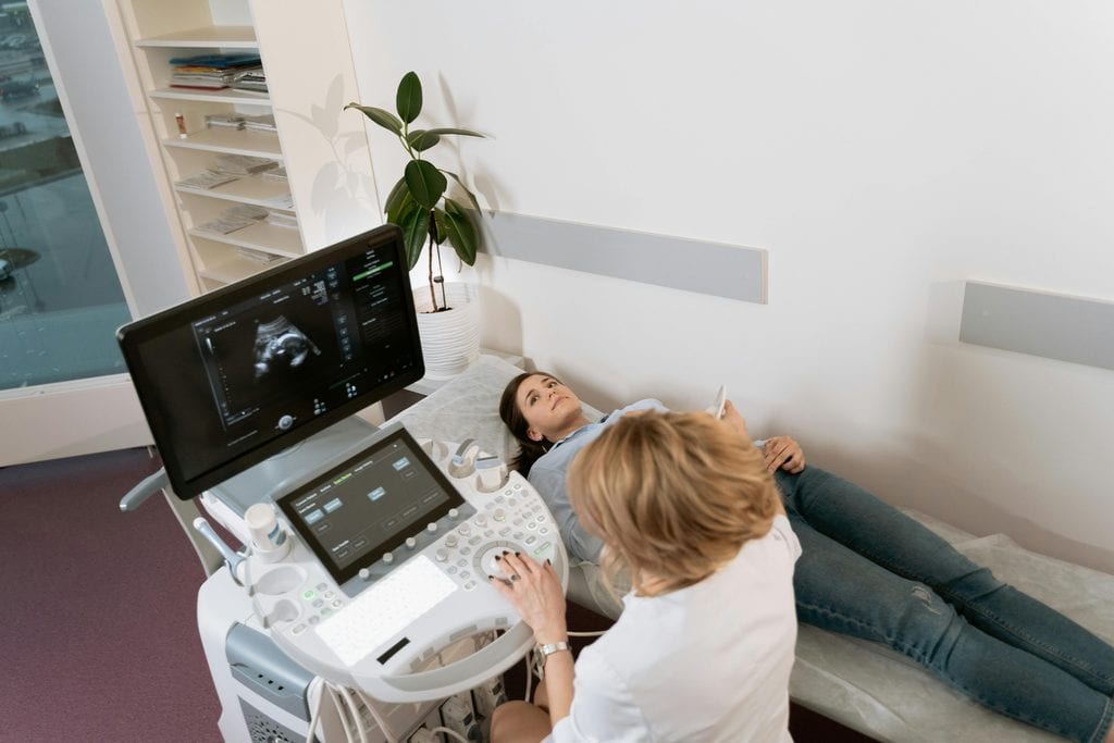
What Does a Gynaecological Ultrasound Scan Examine?
What Does a Gynaecological Ultrasound Scan Examine?
Depending on your symptoms and the type of scan performed, your Sonographer will assess several important pelvic structures.
1. Uterus (Womb)
The scan examines the size, shape, position and appearance of the uterus, looking for structural abnormalities such as fibroids or features suggestive of adenomyosis.
2. Endometrium (Womb Lining)
The endometrium is assessed for its thickness, appearance and any visible irregularities.
This can be particularly important when investigating heavy periods, irregular bleeding or bleeding after menopause.
3. Ovaries
Both ovaries are assessed where they can be visualised, including their size, appearance and the presence of cysts or other abnormalities.
The examination may also identify polycystic ovarian morphology.
4. Adnexal Regions
The areas surrounding the uterus and ovaries are examined for visible masses, cysts or other abnormalities.
5. Pelvic Free Fluid
Your Sonographer may assess for the presence of free fluid within the pelvis, which can sometimes be associated with physiological changes or underlying gynaecological conditions.
6. Other Relevant Pelvic Findings
Depending on the clinical indication, the scan may identify structural changes that warrant further investigation or review by a gynaecologist.
What Is Included in Your Private Gynaecological Ultrasound Appointment?
At Midlands Women’s Health, our ultrasound service is designed to provide a thorough and professional diagnostic assessment.
Your appointment includes:
- Clinical history review: Discussion of your symptoms, menstrual history and reason for requesting the scan.
- Professional Sonographer assessment: Your examination is performed by an experienced, qualified Sonographer.
- Transabdominal pelvic ultrasound: External assessment of the pelvic organs where clinically appropriate.
- Transvaginal pelvic ultrasound: Detailed internal examination where indicated and with your consent.
- Assessment of pelvic structures: Examination of the uterus, endometrium, ovaries and surrounding pelvic areas where visible.
- Explanation of the examination: Your Sonographer will guide you through the process and explain what to expect.
- Ultrasound findings and reporting: A diagnostic ultrasound report documenting the findings.
- Recommendations for further assessment: Where appropriate, findings may indicate the need for specialist gynaecological review or additional investigations.
The examination may involve one or both scanning techniques, depending on clinical suitability and your preferences.
How to Prepare for Your Gynaecological Ultrasound Scan

Preparation depends on whether you are having a transabdominal or transvaginal examination.
Preparing for a Transabdominal (TA) Ultrasound
A full bladder helps improve ultrasound visibility by providing a clearer view of the pelvic organs.
Before your appointment:
- Drink approximately 1 litre of water around one hour before your scan, unless you have been advised to restrict fluids.
- Try not to empty your bladder before your examination.
- Wear comfortable, loose-fitting clothing that allows easy access to your lower abdomen.
- Bring any relevant previous scan reports or medical information, if available.
Your bladder should feel comfortably full rather than painfully full. Please inform your Sonographer if you are uncomfortable.
Preparing for a Transvaginal (TV) Ultrasound
Unlike an abdominal ultrasound, a transvaginal scan is usually performed with an empty bladder.
Before your examination:
- You will usually be asked to empty your bladder immediately before the scan.
- Wear comfortable clothing that is easy to adjust or remove from the waist down.
- Inform your Sonographer if you are pregnant or think you may be pregnant.
- Let your Sonographer know about any pelvic discomfort, previous difficult examinations or concerns about an internal scan.
If both scans are required, the abdominal examination is generally completed first, followed by the transvaginal scan after you have emptied your bladder.
Can I have a pelvic ultrasound during my period?
Yes. A pelvic ultrasound can often be performed during menstruation. However, the optimal timing may depend on the reason for your scan. Our team can advise if a particular stage of your menstrual cycle is preferable.
Why Might I Need a Private Pelvic Ultrasound?
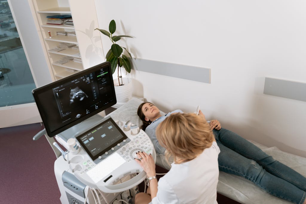
A gynaecological ultrasound may be recommended if you are experiencing:
- Heavy menstrual bleeding or prolonged periods.
- Irregular periods or bleeding between periods.
- Pelvic pain, pressure or discomfort.
- Painful periods (dysmenorrhoea).
- Postmenopausal bleeding.
- Suspected ovarian cysts.
- Suspected uterine fibroids.
- Fertility difficulties or concerns about reproductive health.
- Symptoms associated with PCOS.
- Suspected adenomyosis.
- Pelvic bloating or fullness.
- Follow-up of previously identified gynaecological abnormalities.
An ultrasound can provide important diagnostic information, but it does not detect every gynaecological condition. Further clinical assessment or investigations may sometimes be necessary.
What Happens After My Ultrasound Scan?
Following your examination, your Sonographer will document the findings in a diagnostic ultrasound report.
The report can help your referring clinician, GP or gynaecologist understand the ultrasound findings and determine whether further assessment or treatment is required.
If an abnormality is identified, additional investigations or specialist review may be recommended.
At Midlands Women’s Health, we also offer access to specialist gynaecological consultations, supporting a joined-up approach to the investigation and management of women’s health concerns.
An ultrasound provides valuable diagnostic information but does not replace a full clinical assessment when symptoms persist or require further investigation.
Why Choose Midlands Women’s Health for Imaging?
Experienced Sonographer
Your examination is undertaken by a qualified Sonographer with extensive experience in diagnostic ultrasound and women’s health imaging.
Comprehensive Pelvic Assessment
We offer transabdominal and transvaginal scanning, enabling the most appropriate examination for your symptoms and individual circumstances.
Compassionate, Patient-Centred Care
We understand that gynaecological symptoms and intimate examinations can sometimes feel worrying. We provide a respectful, supportive environment where your comfort and dignity are prioritised.
Specialist Gynaecological Support
Where further assessment is indicated, our wider specialist gynaecology services can support investigation, diagnosis and treatment planning.
Private Women’s Health Services in Birmingham
Midlands Women’s Health provides accessible private gynaecological services for women seeking personalised care and diagnostic assessment.
Helpful Websites & Support Resources – Pelvic Imaging
The following trusted organisations provide reliable information about pelvic imaging, including ultrasound scans, MRI, investigations for pelvic pain, abnormal bleeding, ovarian cysts, fibroids, endometriosis, fertility concerns, and other gynaecological conditions.
| Organisational Name | Link | Description |
| Royal College of Obstetricians and Gynaecologists | https://www.rcog.org.uk/for-the-public/ | Provides evidence-based information about gynaecological conditions and investigations, including the role of imaging in assessing symptoms such as pelvic pain, heavy bleeding, fibroids, ovarian cysts, and fertility concerns. |
| NHS | https://www.nhs.uk/conditions/ultrasound-scan/ | Trusted NHS information explaining how ultrasound scans work, what they are used for, how to prepare, and what patients can expect during the procedure. Pelvic ultrasound is commonly used to assess the womb, ovaries, and surrounding pelvic structures. |
| RadiologyInfo | https://www.radiologyinfo.org/ | Provides patient-friendly information about medical imaging tests, including pelvic ultrasound and MRI scans, explaining why they are performed, how they work, and what patients can expect. |
| Women’s Health Concern | https://www.womens-health-concern.org/ | Provides accessible, evidence-based information on women’s health conditions, symptoms, and investigations, helping patients understand common gynaecological problems and diagnostic pathways. |
Frequently Asked Questions
Most patients experience little or no pain, although some may notice mild pressure or discomfort. If you experience pain, inform your Sonographer immediately. The examination can be adjusted or stopped at any time.
For a transabdominal scan, a comfortably full bladder is usually required. For a transvaginal scan, your bladder should normally be empty.
If both examinations are planned, your Sonographer will guide you through the process.
Can a pelvic ultrasound detect ovarian cysts?
Yes. Pelvic ultrasound is commonly used to identify and assess ovarian cysts, including their size and ultrasound appearance.
Yes. Ultrasound is commonly used to identify uterine fibroids and assess their location and size.
Ultrasound may identify ovarian endometriosis and some features of deep endometriosis, particularly when performed using specialist techniques and by a Consultant Radiologist like Dr Moji Balogun. However, superficial endometriosis may not be visible, and a normal ultrasound does not rule out endometriosis. If you wish to explore a scan with Dr Balogun please access her page under Our Specialists.
Ultrasound can identify polycystic ovarian morphology, but PCOS is diagnosed using a combination of clinical history, symptoms and, where appropriate, blood tests. Ultrasound findings alone do not necessarily confirm PCOS.
Previous sexual activity does not automatically determine whether a transvaginal scan is appropriate. Your Sonographer will discuss the examination, its suitability and alternatives with you. An internal scan will only be performed with your informed consent.
A routine pelvic ultrasound typically takes approximately 30 minutes, although this may vary depending on the examination required and individual findings.
A normal ultrasound does not exclude every gynaecological condition. If you continue to experience pelvic pain, abnormal bleeding or other symptoms, further assessment by a GP or gynaecologist may be appropriate.
Have Questions? Contact Us
Not sure which service is right for you?
Our friendly team at Midlands Women’s Health, a private women’s health clinic in Birmingham, is here to help
Get in Touch With Us
Address
Kat and Co, 20 Calthorpe Road, Edgbaston, Birmingham, B15 1RP
Phone

We provide fast, confidential, and patient-centered care to women across Birmingham and the Midlands
Let us help you take the next step towards better pelvic health, personalised gynaecology care, and improved quality of life.